FIG. 7.
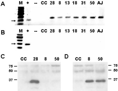
CVB3/TD strains are neutralized by anti-CVB3 polyclonal serum and infect cells to produce viral proteins. (A) Cultures of 105 HeLa cells were inoculated with 103 TCID50 units of CVB3/28 (lane 28), 107 rTCID50 TD8 (lane 8), 104 rTCID50 TD13 (lane 13), 105 rTCID50 TD18 (lane 18), 105 rTCID50 TD31 (lane 31), 106 rTCID50 TD50 (lane 50), or 0.1 ml of heart homogenate taken 18 days p.i. (lane AJ). Total RNA was prepared after 72 h for RT-PCRs with primers KS1 and KS2 (amplimer size, 390 bp). Lane M, 100-bp marker; lane +, CVB3/28 viral RNA RT-PCR; lane -, negative control RT-PCR; lane CC, uninfected cell control. The arrow indicates a 400-bp marker. (B) Same as in panel A, except CVB3 antiserum (1:500) was added to virus stocks in infections, washes, and medium. (C) Cultures of 105 HeLa cells were inoculated with 106 TCID50 units of CVB3/28 (lane 28), 107 rTCID50 TD8 (lane 8), 107 rTCID50 TD50 (lane 50), or uninfected cell control (lane CC). Cells were harvested at 7 h, resuspended in Laemmli buffer, electrophoresed, and analyzed by Western blotting, and viral proteins were detected as described in Materials and Methods. Mobility of molecular weight markers of 150, 75, 50, and 37 is noted. (D) Same as in panel C, but cells were harvested at 26 h p.i.

