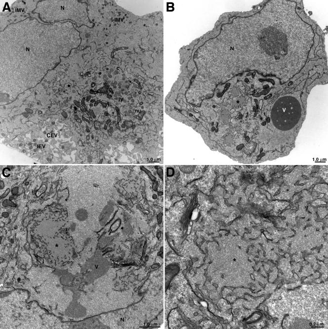
FIG. 8.
Electron microscopy of infected cells. BS-C-1 cells were infected with 3 PFU per cell of vA11Ri in the presence (A) or absence (B to D) of 25 μM IPTG. Cells were fixed and prepared for transmission electron microscopy at 20 h after infection. Electron micrographs are shown with their scale indicated by the bars. Abbreviations: c, crescent; IV, immature virion; nu, nucleoid within an IV; IMV, intracellular mature virion; IEV, intracellular enveloped virion; CEV, cell-associated enveloped virion; V, viroplasm; ✽, intermediate density area; N, nucleus.

