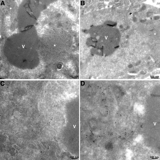
FIG. 9.
Localization of A17, A3 (P4b), D13, and PDI by immunoelectron microscopy. Cells were infected with 3 PFU per cell of vA11Ri in the absence of IPTG. After 24 h, cells were fixed, cryosectioned, and incubated with anti-D13 (A), anti-P4b/4b (B), anti-A17-N (C), or anti-PDI antisera (D), followed by an appropriate secondary antibody and colloidal gold coupled to protein A. Electron micrographs are shown with scale bars. Abbreviations: V, viroplasm; ✽, intermediate density area.

