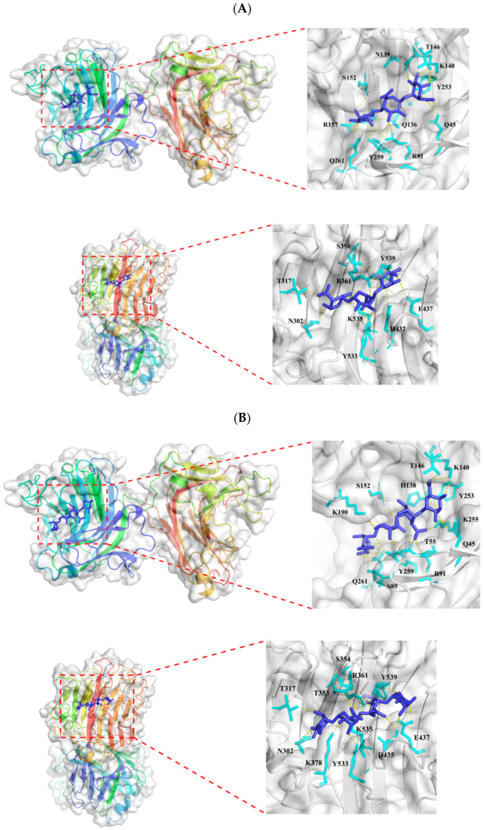
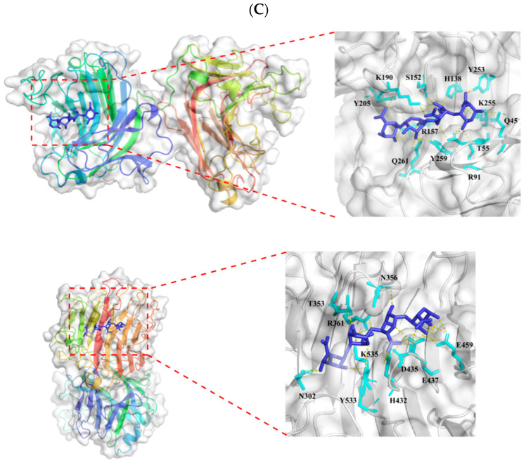
Figure 3.
Molecular docking analysis of Alg169. Binding sites and docked poses of (A) alginate, (B) L-tetraguluronic acid, and (C) D-tetramannuronic acid are shown in the three panels, respectively. For each panel, the upper section shows the docking result for the ligand with Domain 1 of Alg169, and the lower section shows the docking result for the ligand with Domain 2 of Alg169. A detailed view highlighting the residues forming polar contacts with the ligand is shown on the right for each section. The protein structure is colored with a rainbow gradient which varies from the N-terminus (blue) to the C-terminus (red).


