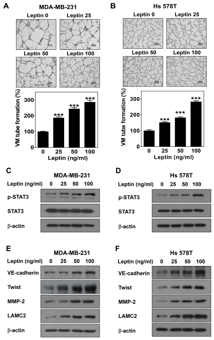
Figure 1.
Leptin promotes VM in human breast cancer cells. VM tube formation assay was carried out in MDA-MB-231 cells (A) and Hs 578T cells (B) treated with leptin. After 16 h of incubation, images were visualized (40× magnification; scale bar = 250 μm) and the number of VM structures formed was counted. Western blot was performed using specific antibody in MDA-MB-231 cells (C,E) and Hs 578T cells (D,F) treated with leptin for 30 min (C,D) or 24 h (E,F). Data are reported as the mean ± SD and statistical significance is indicated as *** p < 0.001 vs. untreated control.

