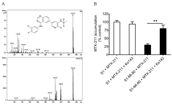
Figure 2.
Intracellular accumulation of MTX-211 (with a molecular weight of 478 g/mol) in cancer cells overexpressing ABCG2. (A) The chemical structure (upper panel) and its fragment ions (lower panel) of MTX-211. (B) The quantification of intracellular MTX-211 concentration using LC-SRM/MS analysis in parental S1 cells (open bars) and ABCG2-overexpressing S1-MI-80 cells (filled bars) in the presence or absence of Ko143, as detailed in the Materials and Methods. The values represent the mean ± S.D. derived from a minimum of three independent experiments. ** p < 0.01, compared to treatment with Ko143.

