FIG. 2.
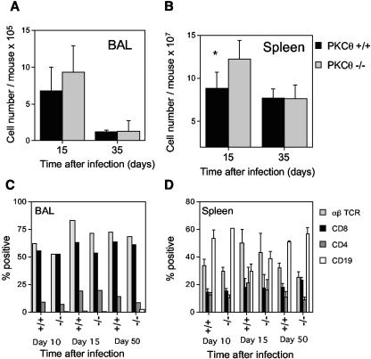
Cell numbers and lymphocyte subsets in the BAL or spleens of PKCθ+/+ and PKCθ−/− mice. The numbers of cells in the BAL (A) or spleen (B) were determined at intervals after intranasal infection of PKCθ+/+ and PKCθ−/− mice with MHV-68. Data are means ± standard deviations (error bars) of cell counts for two separate experiments on cells 15 days after infection and for one experiment on cells 35 days after infection. Cell counts from three or four individual mice were performed at each time point. BAL (C) or spleen cells (D) were stained with phycoerythrin- or fluorescein isothiocyanate-conjugated monoclonal antibodies, and the resulting populations were analyzed by flow cytometry. The means ± standard deviations (error bars) of data from two separate experiments on cells 15 days after infection and one experiment each on cells 10 and 35 days postinfection are shown. Groups of two to four mice were used at each time point. BAL cells were pooled in each experiment, whereas individual spleen cell suspensions were analyzed. The asterisk denotes that there was a statistically significant difference in spleen cell numbers at day 15 after infection in PKCθ−/− and PKCθ+/+ mice (P < 0.05 by Student's t test).

