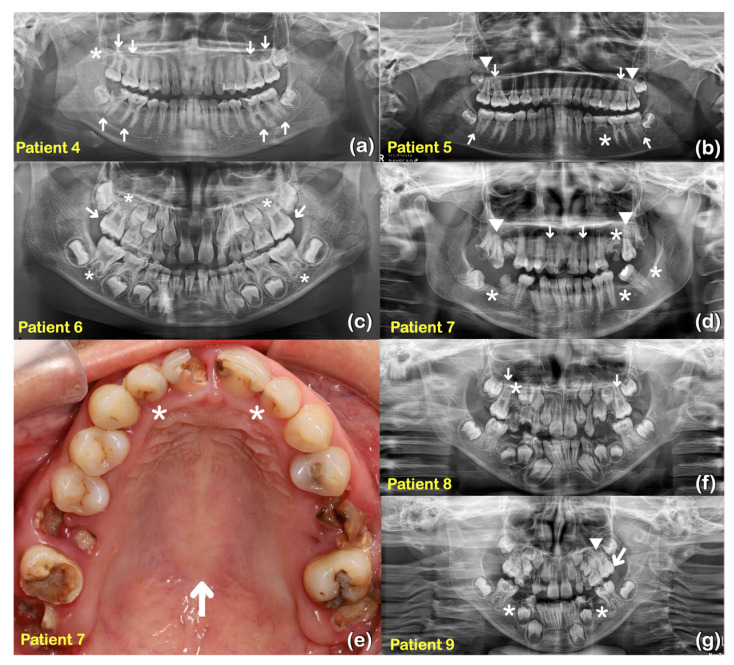
Figure 3.
Family 1 (a) Patient 4 at age 14 years. Panoramic radiograph showing taurodontism of teeth 16, 17, 26, 27, 36, 37, 46, and 47 (arrows). Generalized long roots. Agenesis of tooth 18 (asterisk). (b) Patient 5 at age 12 years. Panoramic radiograph showing taurodontism of teeth 16, 26, 37, and 47 (arrows). Unseparated roots of teeth 17 and 27 (arrowheads). Generalized thin and tapered roots. Agenesis of tooth 35 (asterisk). (c) Patient 6 at age 7 years. Panoramic radiograph showing mixed dentition and taurodontism of teeth 16 and 26 (arrows). Shortened roots of teeth 16, 26, 36, and 46 (asterisks). Family 2 (d) Patient 7 at age 28 years. Panoramic radiograph showing unseparated roots of teeth 17 and 27 (arrowheads). Agenesis of teeth 25, 36, 38, and 47 (asterisks). Microdontia of teeth 12 and 22 (arrows). (e) Patient 7. Microdontia of teeth 12 and 22 (asterisks). Torus palatinus (arrow). (f) Patient 8 at age 10 years. Panoramic radiograph showing mixed dentition. Taurodontism of teeth 16 and 26 (arrows). Agenesis of tooth 15 (asterisk). (g) Patient 9 at age 7 years. Panoramic radiograph showing mixed dentition, taurodontism of tooth 26 (arrow). Agenesis of teeth 35 and 45 (asterisks). A supernumerary maxillary tooth (arrowhead).

