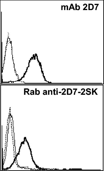
FIG. 7.
Detection of surface CCR5 expression by Rabbit anti-2D7-2SK antibodies. Flow cytometry was used to detect binding of MAb 2D7 and Rab-anti-2D7-2SK to cell surface CCR5 expressed on CEM.NKR.CCR5 cells. Cells were incubated with 25 μg/ml concentrations of each antibody, which were detected with a FITC-labeled goat anti-mouse (top panel) or FITC-labeled goat anti-rabbit IgG (bottom panel). Flow cytometry histograms from a representative experiment are shown: staining of CEM.NKR (thin lines) and CEM.NKR.CCR5 (thick lines) cells with anti-CCR5 MAb 2D7 (top panel) or rabbit anti-2D7-2SK (bottom panel) or with isotype matched controls (dotted lines). Top panel, mouse IgG; bottom panel, preimmune rabbit IgG.

