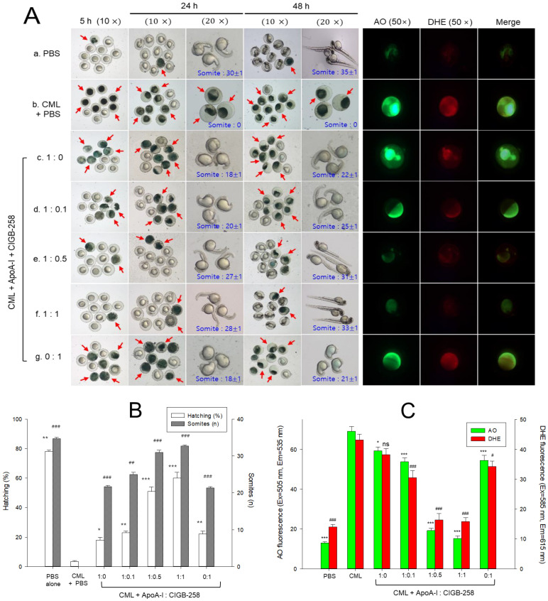
Figure 7.
Effect of different combinations of apolipoprotein A-I (apoA-I) and CIGB-258 on the developmental changes, reactive oxygen species generation (ROS) and apoptosis in carboxymethyl lysine (CML) treated zebrafish embryos. (A) Pictorial view of the developmental changes during 5–48 h post-treatment (the red arrow indicates the dead embryos). ROS and apoptosis levels were determined by dihydroethidium (DHE) and acridine orange (AO) fluorescent staining (images were captured at 5 h post-treatment). (B) Hatching (%) and somites (n) depicting quantification of developmental changes at 24 h post-treatment. (C) Image J-based quantification of AO and DHE fluorescent intensities. PBS and CML+PBS group received 10 nL microinjection of PBS and 500 ng CML in PBS, respectively; apoA-I:CIGB-258 (1:0) and apoA-I:CIGB-258 (0:1) group injected with 1.4 ng/10 nL apoA-I, and 143 pg/10 nL CIGB-25, respectively; apoA-I:CIGB-258 (1:0.1) or (1:0.5) or (1:1) injected with 10 nL of 1.4 ng apoA-I containing 14 pg or 70 pg or 143 pg for CIGB-258. Statistical significance denoted by *, ** and *** at p < 0.05, p < 0.01 and p < 0.001 for AO fluorescent and hatching (%); #, ##, and ### at p < 0.05, p < 0.01 and p < 0.001 for DHE fluorescent and somite counts were compared to the CML+PBS group, respectively; ns represent nonsignificant difference between the groups.

