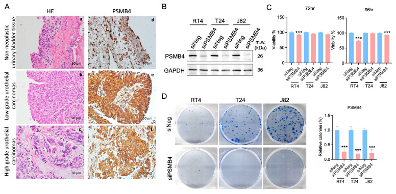
Figure 1.
PSMB4 expression in patients with bladder cancer and bladder cancer cell lines. (A) a–c: Nonneoplastic urinary bladder tissue; low-grade and high-grade urothelial carcinoma tissues (H&E, 400×). d–f: Immunohistochemical staining of PSMB4 in nonneoplastic urinary bladder tissue and in low-grade and high-grade urothelial carcinoma tissues (PSMB4, 400×). Scale bar = 50 μm. (B) PSMB4 protein expression after siPSMB4 transfection for 72 h in RT4, T24, and J82 cells (n = 10). (C) The viability of RT4, T24, and J82 human bladder cancer cells was evaluated using an MTT assay after PSMB4 silencing for 72 and 96 h (n = 6). (D) Colony formation assay of bladder cancer cells after PSMB4 downregulation (n = 5). Data are presented as the mean ± SEM. *** p < 0.001 compared to the siRNA negative control group, determined by unpaired t-test with the Mann–Whitney test.

