Table 2.
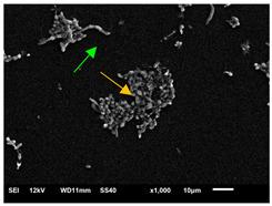
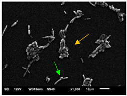
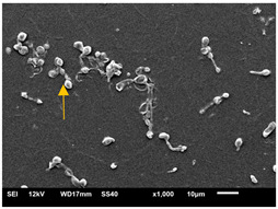
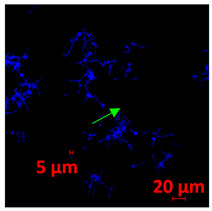
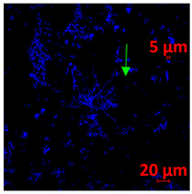
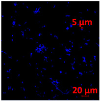
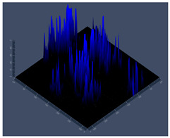
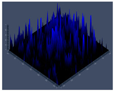
Scanning electron microscopy (SEM) images and confocal microscopy (CLSM) images in two-dimensional and three-dimensional views of strain EH-315 of H. capsulatum after the formation of the initial biofilm (24 h) (A–I) and mature biofilm (144 h) (J–R) in HAM-F12 medium (control), HAM-F12 + 20 µM zinc, and HAM-F12 + 10 µM TPEN. All representative images were magnified 1000× (10 µM). Green arrows—yeast/yellow arrows—hyphae.
| EH-315 | |||
|---|---|---|---|
| Time | HAM-F12 Control | HAM-F12 + 20 µM Zinc | HAM-F12 + 10 µM TPEN |
| 24 h SEM |
A
|
B
|
C
|
| 24 h CLSM 2D |
D
|
E
|
F
|
| 24 h CLSM 2.5D |
G
|
H
|
I
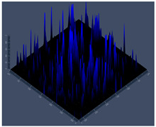
|
| 144 h SEM |
J
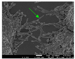
|
K
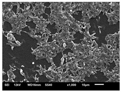
|
L
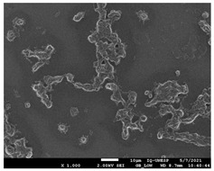
|
| 144 h CLSM 2D |
M
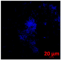
|
N
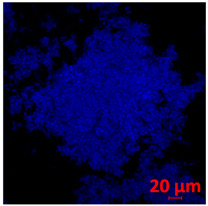
|
O
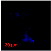
|
| 144 h CLSM 2.5 D |
P
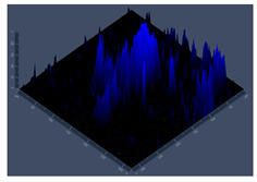
|
Q
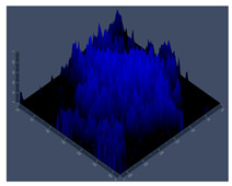
|
R
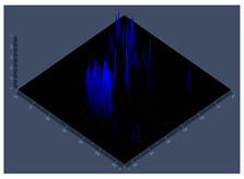
|
