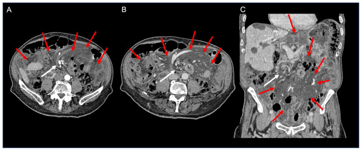
Figure 2.
Contrast-enhanced CT axial arterial (A) and venous phase (B) images and coronal venous phase (C) image of the abdomen showing diffuse fat stranding of the mesentery (red arrows), containing multiple nodes. Note the whirl-like rotation of the mesentery and mesenteric veins around the superior mesenteric artery (“whirlpool sign”—white arrows).

