Figure 5.
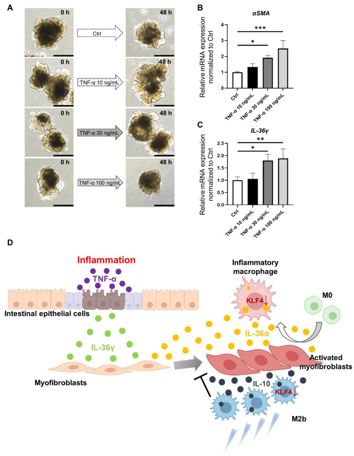
Analysis in an ex vivo model reconstituting IBD using HIOs. (A). Macroscopic images of organoids stimulated with TNF-α. Scale bars: 500 µm. (B). Relative expression of αSMA mRNA in control organoids (without TNF-α addition) and organoids supplemented with TNF-α at 30 ng/mL and 100 ng/mL. Relative expression of αSMA mRNA was significantly increased in the TNF-α 30 ng/mL and TNF-α 100 ng/mL groups compared with the control (p < 0.05 and p < 0.001, respectively); mean ± SD; n = 3. Statistical analysis was performed using two-tailed Dunnett’s test; * p < 0.05, *** p < 0.001. (C). Relative expression of IL-36γ mRNA in control organoid and organoids supplemented with TNF-α at each concentration. Relative expression of IL-36γ mRNA was significantly increased in the 30 ng/mL and 100 ng/mL groups compared with the control (p < 0.05 and p < 0.01, respectively); mean ± SD; n = 3. Statistical analysis was performed using two-tailed Dunnett’s test; * p < 0.05, ** p < 0.01. (D). Interactions between the small intestinal epithelium, intestinal stromal myofibroblasts, and macrophages and cytokines involved in intestinal inflammation and fibrosis. Ctrl: control, HIOs: human-induced pluripotent stem cell-derived small intestinal organoids, IBD: inflammatory bowel disease.

