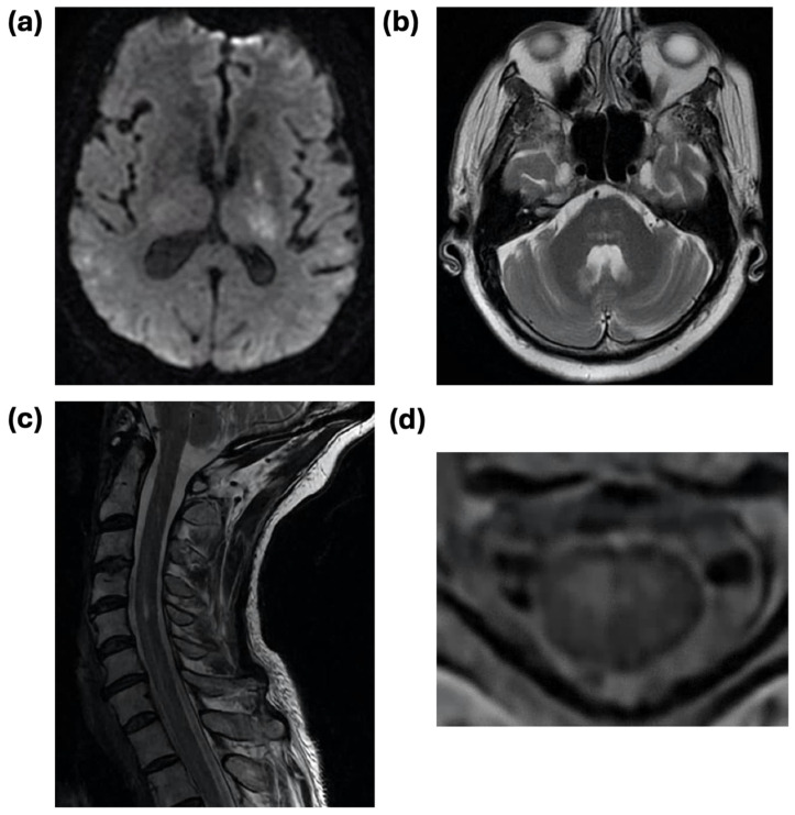
Figure 2.
Representative radiographic features of WNV encephalitis and acute flaccid paralysis. (a) Diffusion-weighted imaging (DWI) axial MRI sequence demonstrating areas of restricted diffusion in the bilateral thalami with associated T2/FLAIR signal in an 83-year-old man with fatal WNV encephalitis who presented with dizziness, slurred speech, and falls. (b) T2/FLAIR axial MRI sequence demonstrating patchy bilateral signal in the pons in a 69-year-old man presenting with altered mental status and diagnosed with WNV encephalitis. (c) Sagittal T2 MRI sequence demonstrating hyperintensity of the cervical spinal cord extending from C3-C4 to the thoracic cord in a 57-year-old woman who presented with greater right than left proximal weakness progressing to respiratory failure, diagnosed with WNV acute flaccid paralysis. (d) Axial T2 MRI sequence of the patient in (c) demonstrating right greater than left cord hyperintensity, particularly of the ventral cord.

