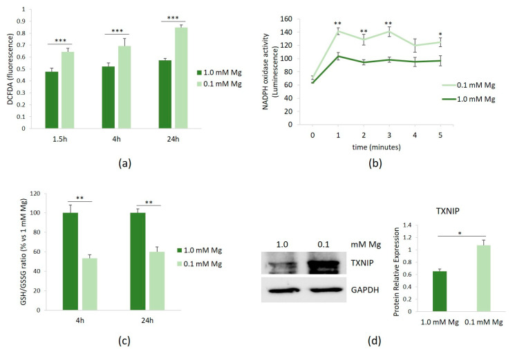
Figure 1.
ROS in HUVECs cultured in low Mg. HUVECs were cultured in 0.1 or 1.0 mM Mg-containing medium. All the experiments were performed in triplicate at least three times. (a) After 1.5, 4, and 24 h of culture in media containing 0.1 or 1.0 mM Mg, ROS were measured as described in the methods. (b) After 1.5 h of culture in media containing 0.1 or 1.0 mM Mg, NADPH oxidase activity was measured using a lucigenin-derived chemiluminescence assay. (c) The GSH/GSSH ratio was measured using a luminescence-based assay. The data were expressed as the percentage vs. 1.0 mM Mg ± SD. (d) The total amounts of TXNIP were evaluated by Western blot after 20 h of exposure to low-Mg medium. Anti-GAPDH antibodies were used as a control of equal loading. A representative blot and densitometry performed on three independent experiments and obtained by ImageJ are shown. * p ≤ 0.05; ** p ≤ 0.01; and *** p ≤ 0.001.

