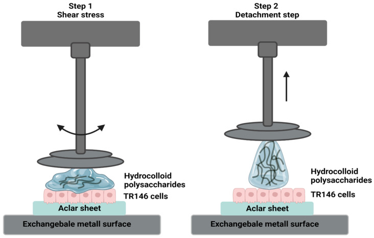
Figure 1.
Schematic illustration of the applied in vitro adhesion setup. In step 1, shear stresses of 0.1 to 100 rad/s are applied to simulate physiological shear rates in the oral cavity. In step 2, the system detaches the aqueous solution containing hydrocolloid polysaccharides from the TR146 cell layer.

