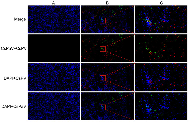
Figure 5.
Detection of the mRNAs of CsPaV and CsPV in the infected spleen of Chinese tongue soles using FISH. (A) Uninfected spleen (Bar, 50 μm); (B) Infected spleen (Bar, 50 μm); (C) High magnification of the red rectangular region (Bar, 10 μm). Tissue sections display red fluorescence for CsPV (Cy3-conjugated probes), green for CsPaV (FAM-conjugated probes), and blue for cell nucleus (DAPI). The mRNAs of CsPV and CsPaV were detected in cytoplasm, which showed red (red arrow) and green (green arrow), and the nucleus showed purple (purple arrow) and cyan (cyan arrow). The mRNAs of the two viruses co-located in the cytoplasm showed yellow (yellow arrow), and the nucleus showed white (white arrow) in one cell.

