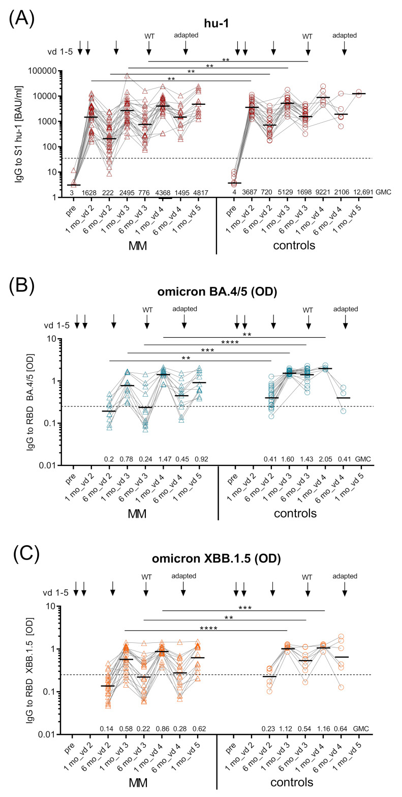
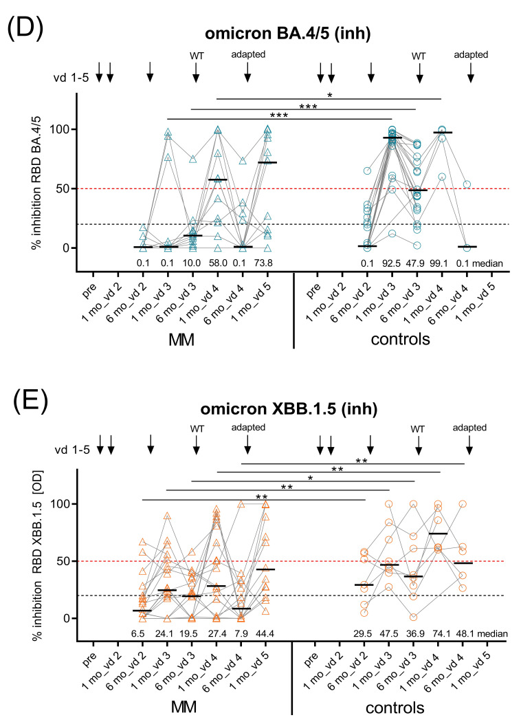
Figure 3.
Kinetics of SARS-CoV-2 spike (S1)-specific IgG antibodies. Kinetics of GMC of (A) ancestral virus hu-1 S1-specific IgG (BAUs/mL) in uninfected MM patients (n = 32) and controls (n = 23) measured before vd1, one and six months after vd2, vd3, and vd4, and one month after vd5 of SARS-CoV-2 mRNA vaccine (BNT162b2 or mRNA-1273); dashed line—positive cut-off for S1-specific IgG at 35.2 BAUs/mL; (B) Omicron BA.4/5 RBD-specific IgG (as OD) in uninfected MM patients (n = 11) and controls (n = 20) six months after vd2, one and six months after vd3 and vd4, and one month after vd5 and C) Omicron XBB.1.5 RBD-specific IgG (as OD) in uninfected MM patients (n = 22) and controls (n = 7) at the same time points; black dashed lines in (B,C), OD > 0.25 considered positive, horizontal line indicates GMC provided numerically above x-axis; kinetics of inhibition capacity of (D) Omicron BA.4/5 RBD-specific IgG (as % inhibition) measured in uninfected MM patients (n = 11) and controls (n = 20) six months after vd2, one and six months after vd3 and vd4, and one month after vd5, and (E) Omicron XBB.1.5 RBD-specific IgG (as % inhibition) measured in uninfected MM patients (n = 22) and controls (n = 7) at the same time points; inhibition levels >20% considered positive (black dashed line), inhibition levels >50% relevant (red dashed line); horizontal line indicates median provided numerically above x-axis. Abbreviations: BAUs, binding antibody units; GMC, geometric mean concentrations; IgG, immunoglobulin G; mo, months; MM, multiple myeloma patients; mRNA, messenger ribonucleic acid; OD, optical density; RBD, receptor-binding domain; S1, SARS-CoV-2 spike protein 1; vd, vaccine dose. Linear contrasts with Sidak–Holm-corrected p-values; **** p ≤ 0.0001; *** p ≤ 0.001; ** p ≤ 0.01; * p ≤ 0.05.


