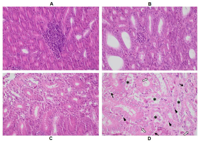
Figure A1.
Results of histological examination: (A) marked focal mononuclear cell infiltration in the interstitium (H-E; 400X); (B) mild mononuclear cell infiltration in the interstitium surrounded with normal tubuli (H-E; 400X); (C) multifocal tubular damage with marked cellular detachment (H-E; 400X); (D) marked tubular lesions in the kidney with cellular detachment (black arrow), hydropic degeneration (white arrow), and karyopyknosis (arrowhead). Detached cells and cellular debris can be seen in multiple tubuli (asterisk)—H-E; 600X.

