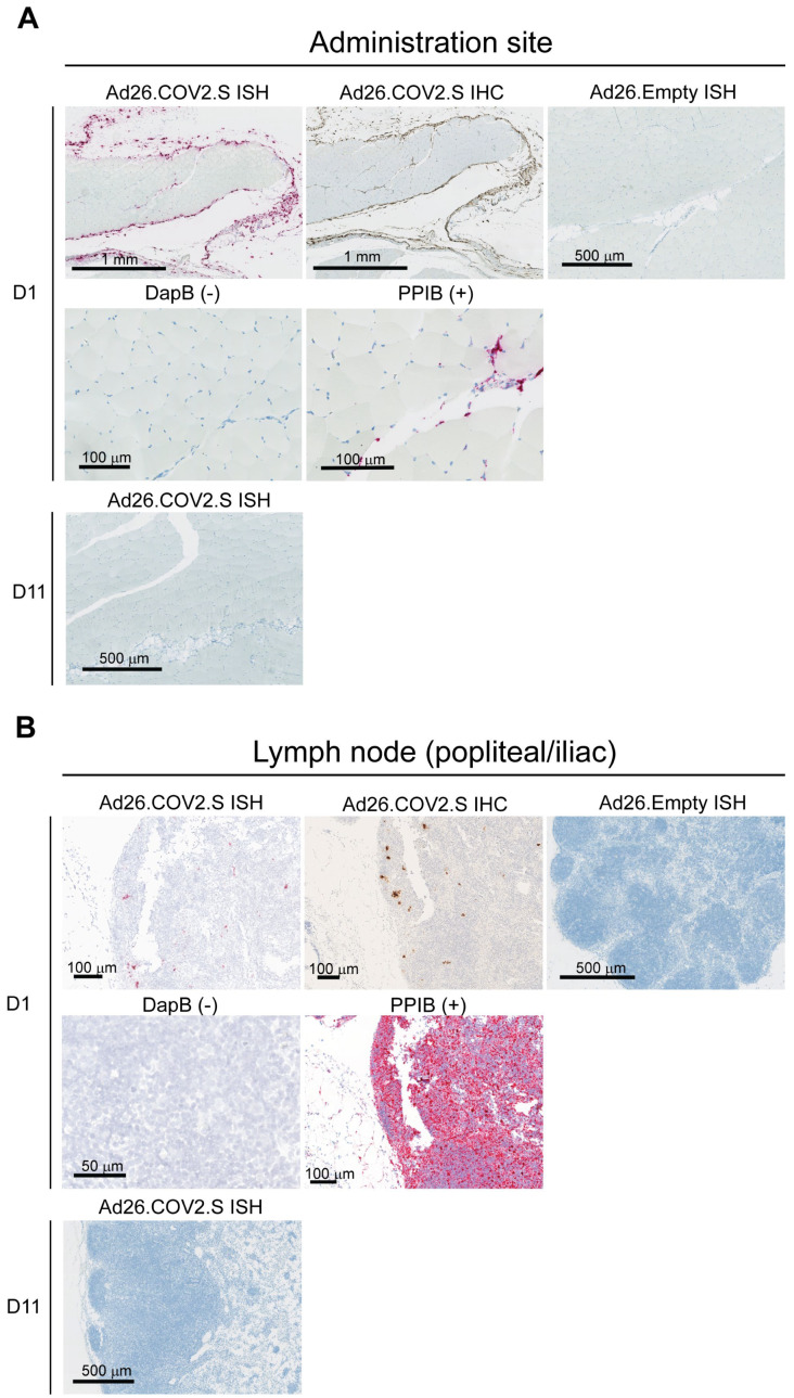
Figure 2.
Spike mRNA in situ hybridization (ISH) detected at site of injection and draining lymph nodes 1 day after IM Ad26.COV2.S dosing in rabbits. Anti-SARS-CoV2 S1 staining by immunohistochemistry (IHC) and ISH of spike mRNA at (A) the administration site and in (B) lymph nodes from rabbits (n = 4 per group) at Day 1 after dosing with Ad26.COV2.S (positive) or Ad26.Empty (negative) or ISH on Day 11 after dosing with Ad26.COV2.S (negative). The bacterial gene dihydrodipicolinate reductase (dapB) (negative control) and housekeeping gene cyclosporine-binding protein peptidylpropyl isomerase B (PPIB) (positive control) are shown in the same tissues. In brown (IHC) are the positive areas for the target protein and in purple/red are the positive areas for the target mRNA (ISH). The black bar represents the magnification of the image.

