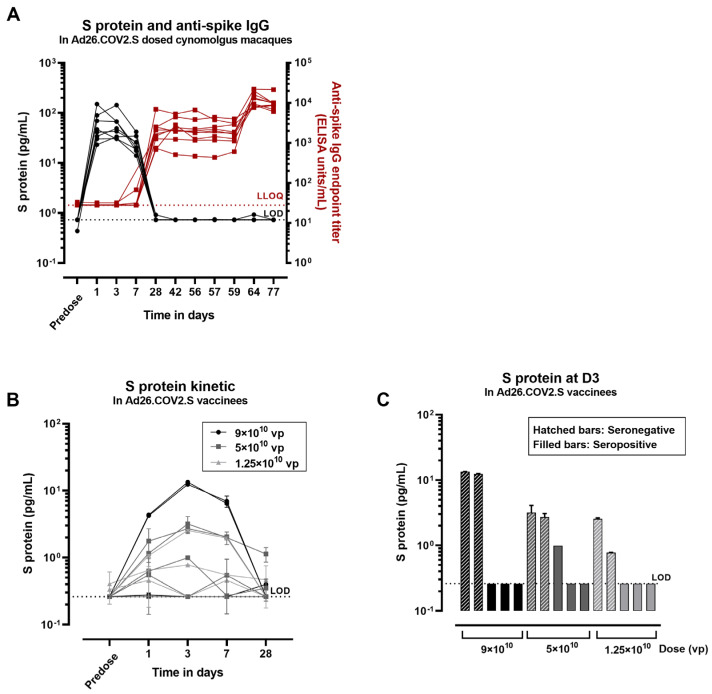
Figure 6.
Concentration of S protein in serum after dosing with Ad26.COV2.S in macaques and humans. (A) Macaque serum samples were analyzed for S-protein detection and for anti-spike immunoglobulin G (IgG) antibody titers. Black symbols correspond to S concentration expressed in pg/mL and red symbols correspond to anti-spike IgG titers expressed as endpoint titer ELISA (1 symbol/animal). The black dotted line represents the lower limit of detection (LOD) of the S assay based on the standard curve. The red dotted line corresponds to the lower limit of quantification of the anti-spike assay (LLOQ). (B) The concentration of S protein was measured in Ad26.COV2.S vaccinees at different timepoints before and after dosing. (C) The concentration of the S protein was measured in serum from seropositive vaccinees (containing anti-spike neutralizing antibodies) or seronegative vaccinees 3 days after dosing with Ad26.COV.2.S. The black dotted line represents the lower limit of detection (LOD) of the assay based on the standard curve. The error bars represent the standard deviation of 2 technical replicates in (B,C).

