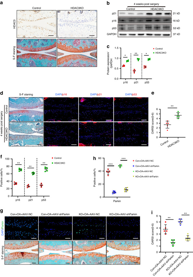
Fig. 4.
Loss of HDAC3 activates PINK1/Parkin signaling to promote chondrocyte senescence and OA progression. a Representative images of IHC and safranin O/fast green staining of HDAC3 in articular cartilage of HDAC3KO and Control mice at 3 months old. Scale bar: 50 µm. b, c Western Blotting analysis of p16INK4a, p21, p53 in the cartilage of HDAC3KO and Control mice after DMM surgery (n = 5). d Representative images of safranin O/fast green and immunofluorescence staining of p16INK4a, p21, and p53 in HDAC3KO and Control cartilage of mice after DMM surgery. Scale bars: 50 µm. e Quantification of the OARSI scale based on staining results in (d) (n = 5). f Quantification of p16INK4a, p21, and p53-positive chondrocytes based on staining results in (d) (n = 5). g Representative images of safranin O/fast green and immunofluorescence staining of Parkin in HDAC3KO and Control cartilage of mice which intra-articularly injected with AAV-NC or AAV-shParkin after DMM surgery. Scale bar: 50 µm. h Quantification of Parkin-positive chondrocytes based on staining results in (g) (n = 5). i Quantification of the OARSI scale based on staining results in (g) (n = 5). *P < 0.05, **P < 0.01. DMM destablization of the medial meniscus, DAPI 4’,6-diamidino-2-phenylindole, OARSI Osteoarthritis Research Society International, KO knockout, AAV-shParkin adenovirus expressing small hairpin Parkin, AAV-NC negative control

