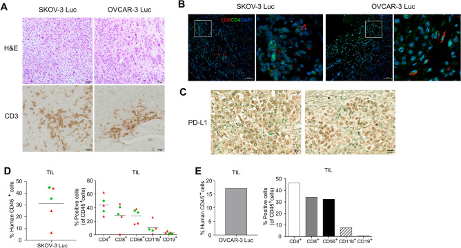
Figure 2.
Presence of tumor-infiltrating lymphocytes (TILs) and immunophenotypical characterization of tumors in Hu-mice injected with ovarian cancer cells. The infiltration of human lymphocytes and the immunophenotype within the ovarian xenograft tumors of Hu-mice were determined by colorimetric immunostaining and flow cytometry analysis. Tumor samples were collected from the peritoneal cavity, processed through formalin fixation and paraffin embedding, then sectioned for histologic examination and analysis of immune cells and PD-L1 expression. (A) The tumor characteristics are visualized via hematoxylin and eosin staining (H&E, top panels) and immunohistochemical staining of CD3+ T cells (bottom panels) in tumors. (B) Immunofluorescence imaging enabled the detection of CD4+ and CD8+ T cells by utilizing anti-human CD4 (Green) and CD8 (Red) antibodies. DAPI was used for nuclear staining (Blue). Magnification, ×20; Scale bar, 50 µm. (C) The expression of PD-L1 expression in SKOV-3 Luc (left panel) and OVCAR-3 Luc (right panel) tumors was determined by an immunohistochemical method using the anti-human PD-L1 antibody (Magnification, ×20; Scale bar, 20 µm). Solid tumor tissues from Hu-mice injected with ovarian cancer cells were dissociated into single cells using enzymes and mechanical methods. These collected cells were then labeled with fluorochrome-conjugated anti-human antibodies. The presence and frequency of human CD45+ cells, CD4+ T cells, CD8+ T cells, CD56+ natural killer (NK) cells, CD11b+ myeloid cells, and CD19+ B cells within the tumor tissues of (D) SKOV-3 Luc (n = 5) and (E) OVCAR-3 Luc (n = 1) models were evaluated using flow cytometry. The first batch was denoted by red triangles, and the second batch was denoted by green circles.

