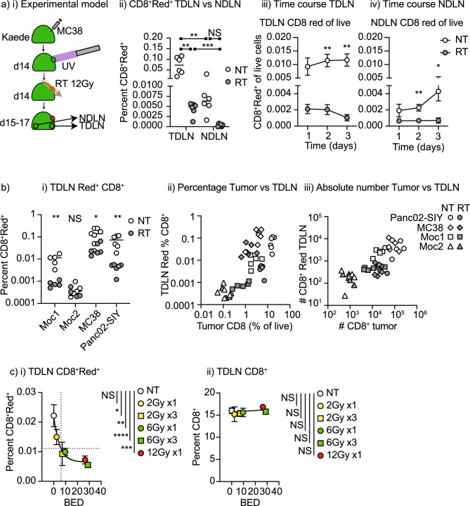
Figure 2.
The effect of tumor radiation therapy on recirculating CD8 T cells in lymph nodes. (a) (i) Kaede mice were implanted with MC38 tumors and at d14 tumors were selectively photoconverted with UV light. Tumors were irradiated with CT-guided radiation to the tumor using a SARRP following photoconversion. Tumors, TdLN and NdLN were harvested over time. (ii) Identification of photoconverted Kaede Red cells in the TdLN, and NdLN 24 h following photoconversion in untreated tumors or tumors that are treated with 12 Gy radiation. Percent of (iii) TdLN and (iv) NdLN cells that are photoconverted over time. (b) (i) Summary of the proportion of photoconverted CD8 T cells in the TdLN of Moc1, Moc2, MC38, and Panc02-SIY tumors when left untreated or treated with 12 Gy radiation to the tumor on a log scale. (ii) Correlation between the photoconverted CD8 T cells in the TdLN and the percent CD8 T cell infiltration of the tumors on a log scale. Open symbols are untreated tumors, closed symbols are tumors treated with 12 Gy radiation. (iii) Data from (ii) represented as absolute numbers in the TdLN or tumor on a log scale. (c) The impact of radiation dose and fractionation on photoconverted CD8 T cells in the TdLN of MC38 tumors. Graphs show (i) the percent photoconverted CD8 T cells in the TdLN or (ii) the percent unconverted CD8 T cells in the TdLN 1d following the last dose of radiation and photoconversion. The x-axis shows BED10 for each dose or dose series. Dotted lines show half maximal photoconverted CD8+ T cells in the TdLN and corresponding BED10. Key. NS not significant; *p < 0.05; **p < 0.01; ***p < 0.001; ****p < 0.0001.

