Fig. 1.

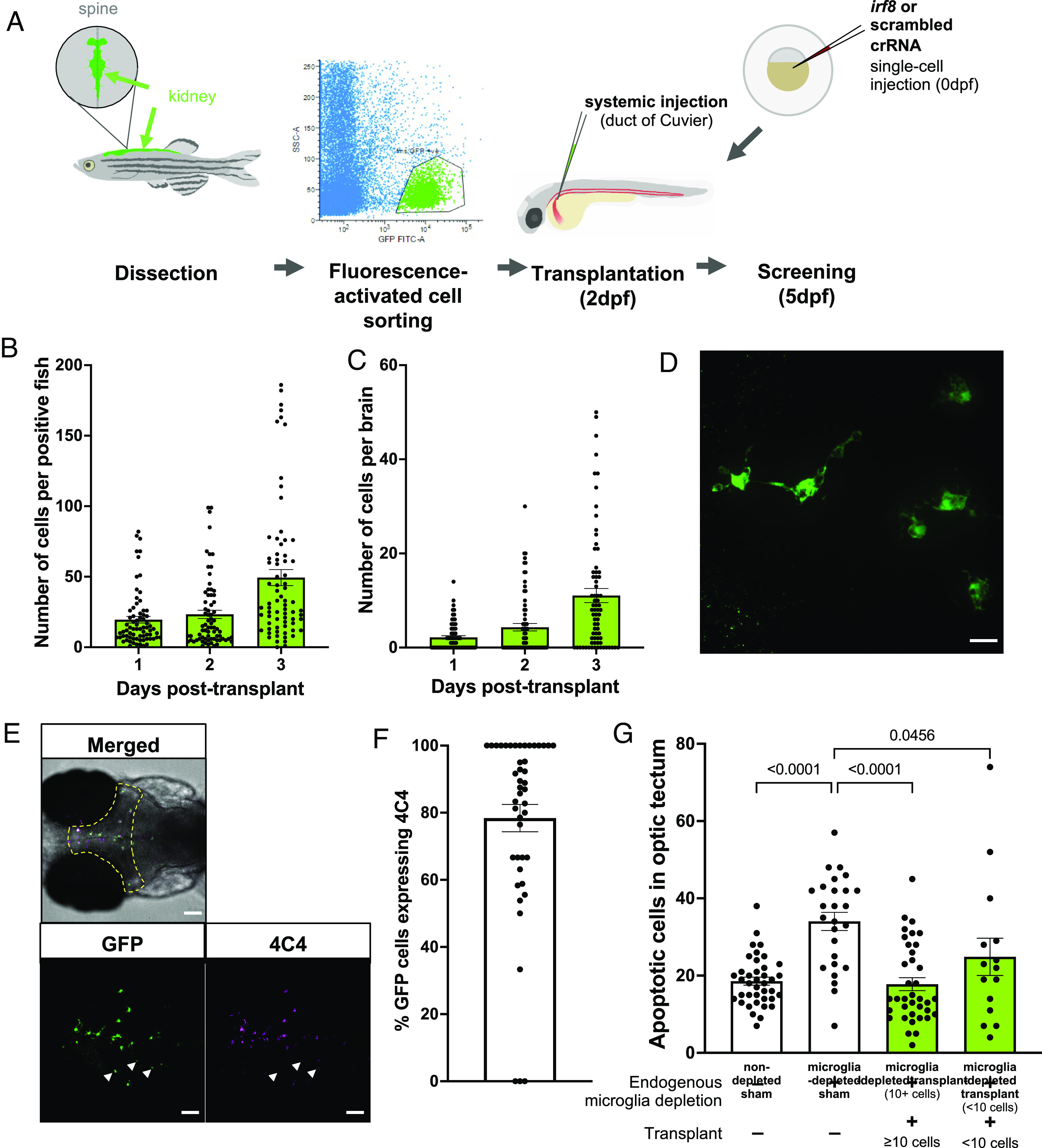
Macrophage transplantation successfully replaces microglia in WT zebrafish. (A) Transplants were performed by dissecting kidneys from Tg(fms:GFP) adult donors and isolating GFP-positive cells using fluorescence-activated cell sorting. Cells were injected into 2 dpf Tg(mpeg1:mCherryCAAX)sh378 hosts via duct of Cuvier. Hosts had previously undergone microglia depletion via targeting of irf8 by CRISPR/Cas9 or were nondepleted with a scrambled control. (B and C) The number of fms:GFP cells per successfully transplanted animal increased throughout the whole body (B) and within the brain (C) across 3 d post-transplant, as revealed by manual counting. (D) Confocal imaging reveals that transplant-derived cells show a branched, microglia-like morphology. The scale bar represents 17 µm. (E and F) Transplant-derived cells express the microglia-specific marker 4C4 at 3 d post-transplant (5 dpf). Colocalization was assessed by manual counting. Arrows indicate GFP-positive cells that do not co-localize with 4C4. The yellow dashed line indicates the border of the optic tectum (region of interest). Three biological replicates, n = 45. The scale bar represents 75 µm. (G) TUNEL staining reveals that transplantation can rescue the number of uncleared apoptotic cells in microglia-depleted brains in a dose-dependent manner, as revealed by manual counting of cells within optic tectum as in E. Kruskal–Wallis test with Dunn’s multiple comparisons. Four biological replicates, n = 15 to 39.
