
Fig. 3.
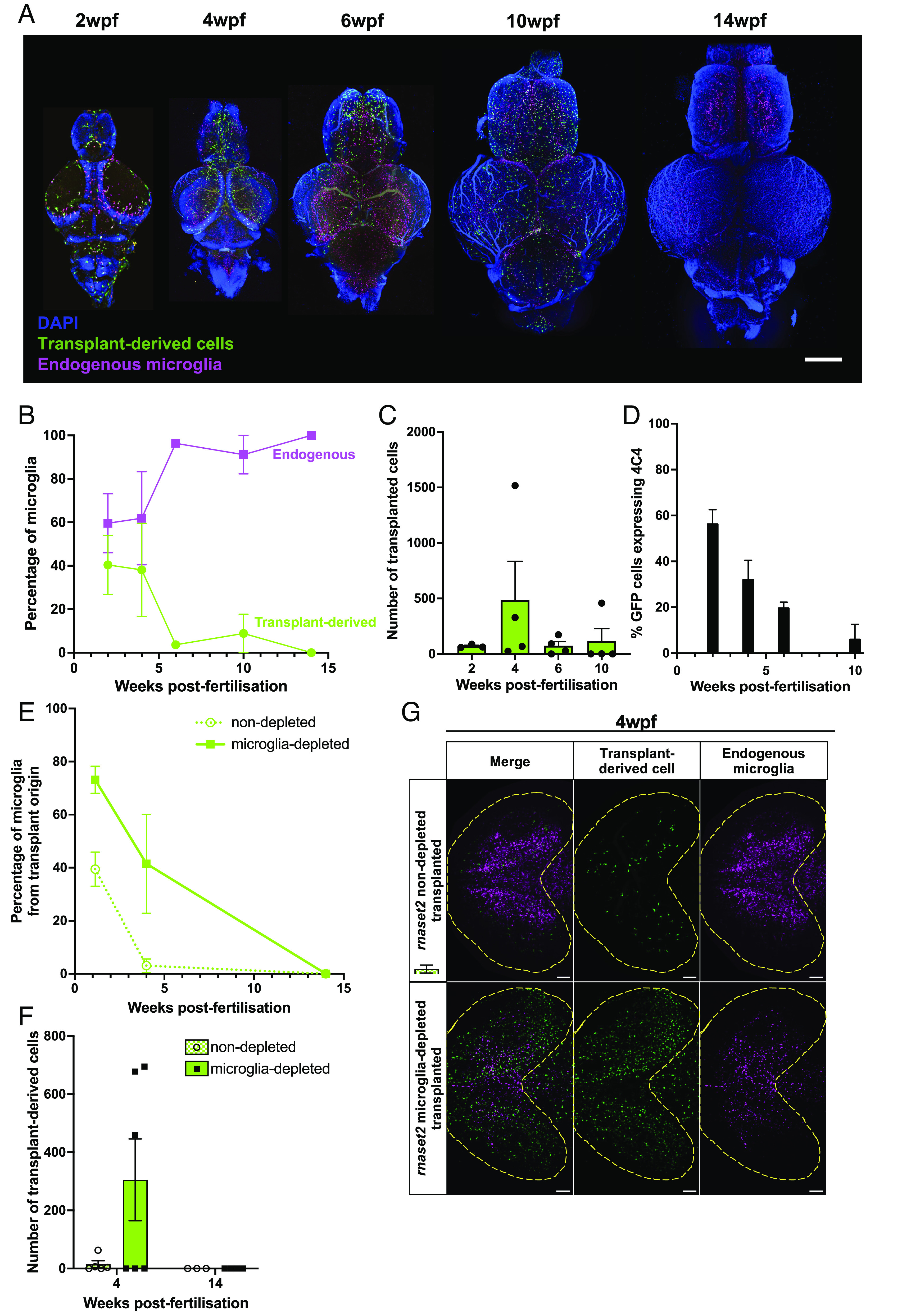
Transplanted cells persist in host brains throughout juvenile stages. (A) Tissue clearing and immunohistochemistry reveal that transplant-derived cells persist in WT microglia-depleted host brains until 10 wpf but are cleared by 14 wpf. Scale bar represents 400 µm. (B) Percentage of microglia from transplant origin gradually decreases in host brains until 14 wpf. (C) The number of transplant-derived cells peaks at 4 wpf and rapidly decreases thereafter. (D) Percentage of transplant-derived cells expressing microglia marker 4C4 steadily decreases from 2 wpf. Three to four biological replicates per timepoint. (E and F) Tissue clearing and immunohistochemistry reveal reduced numbers of transplant-derived cells in nondepleted rnaset2 animals compared with microglia-depleted siblings. Cells are no longer visible in both host groups by 14 wpf. (G) Representative image of transplant-derived cell engraftment at 4 wpf in nondepleted and microglia-depleted rnaset2 hosts. Yellow line indicates optic tectum outline. The scale bar represents 100 µm.