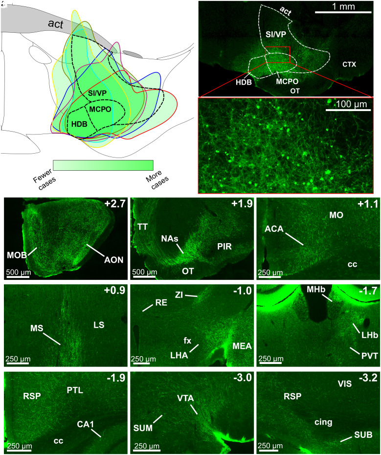
Fig. 3.
BF Npas1+ neurons project to brain areas involved in sleep–wake control, motivated behavior, and olfaction. (A) Composite schematic showing the boundaries of unilateral AAV5-DIO-ChR2-EYFP infusions in the medial BF in six mice, with darker green indicating more frequent transduction of the region. Transduced neurons were largely restricted to the BF. (B) Low (Top) and high (Bottom) magnification images of an individual injection. Abbreviations: act, anterior commissure temporal limb; CTX, cortex; OT, olfactory tubercle. (C–K) Rostral to caudal images of regions with prominent fiber projections. Top Right indicates position relative to bregma. (C and D) BF Npas1+ cells strongly project to olfactory regions, including the main olfactory bulb (MOB) and anterior olfactory nucleus (AON), taenia tecta (TT), and piriform cortex (PIR). (D) BF Npas1+ fibers densely innervate the nucleus accumbens shell (NAs). (E) In the neocortex, BF Npas1+ fibers innervated the anterior cingulate area (ACA) and motor cortex (MO) dorsal to the corpus callosum (cc) and (I and K) the retrosplenial cortex (RS). (F) BF Npas1+ fibers are denser in the medial septum (MS) compared to the lateral septum (LS). (G) Hypothalamic projections were strongest in the lateral hypothalamic area (LHA) close to the fornix (fx) with moderate projections to the zona incerta (ZI). Dense projections were also observed in the amygdala, including the medial amygdalar nucleus (MEA). RE, nucleus reuniens of the thalamus. (H) Projections to the thalamus were densest in the lateral habenula (LHb), dorsal part of the medial habenula (MHb) and paraventricular nucleus (PVT). (H, I, and K) Fibers were present throughout most of the hippocampal formation, including the cornu ammonis 1 (CA1) region, the retrosplenial cortex (RSP), and the subiculum (SUB). Additional abbreviations: Cing, cingulum bundle; PTL, parietal cortex; VIS, visual cortex. (J) Densest midbrain projections were to the ventral tegmental area (VTA) neighboring the supramammillary nucleus (SUM).

