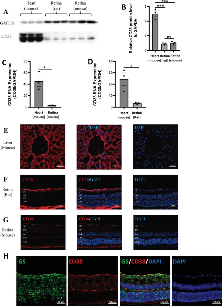
Figure 1.
Expression and localization of CD38 in WT rat and mouse retinal tissues. (A) Detection of CD38 protein expression in mouse heart, rat retina, and mouse retina by WB. (B) Quantitative analysis of CD38 protein expression. (C) Quantitative analysis of CD38 mRNA expression in mouse retina. (D) Quantification of CD38 mRNA expression in rat retina. (E) Localization of CD38 in mouse liver by immunofluorescence. (F) Localization of CD38 in rat retina by immunofluorescence. (G) Localization of CD38 in mouse retina detected by immunofluorescence. (H) Cellular localization of CD38 in the mouse retina determined by GS and CD38 immunofluorescence double staining. GCL, ganglion cell layer; IPL, inner plexiform layer; INL, inner nuclear layer; OPL, outer plexiform layer; ONL, outer nuclear layer; WT, wild type; GS, marker for Müller cells; data presented as: mean ± SEM; ns, not significant; *P < 0.05; ***P < 0.001.

