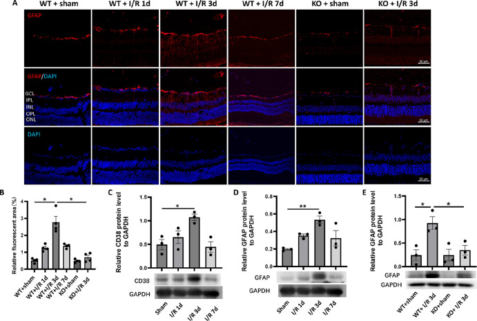
Figure 3.
Effect of CD38 KO on retinal glial activation in mice in the I/R model. (A) GFAP immunofluorescence staining of paraffin sections of mouse retinas. (B) Quantitative statistics of GFAP immunofluorescence staining of paraffin sections of mouse retina. (C) The expression of CD38 protein in retina was detected by WB in sham operation group and I/R on day 1, day 3, and day 7. (D) The expression of GFAP proteins in retina was detected by WB in the sham operation group and I/R on day 1, day 3, and day 7. (E) WB was used to detect changes in CD38 protein expression in retinas at 3 days of I/R in WT and KO mice. GCL, ganglion cell layer; IPL, inner plexiform layer; INL, inner nuclear layer; OPL, outer plexiform layer; ONL, outer nuclear layer. Scale bars = 50 µm; WT, wild type; KO, knockout; data presented as mean ± SEM. *P < 0.05, **P < 0.01.

