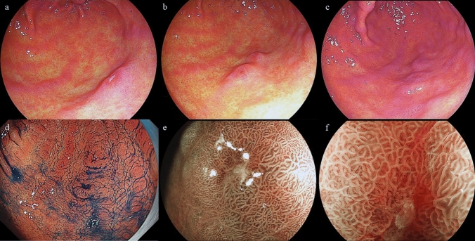
Fig. 1.
a An 8 mm poorly demarcated reddish elevated verrucous gastritis-like lesion under linked color imaging (LCI) located at the greater curvature of the gastric antrum. b This lesion changed to a two-hump shape after 1 year. c Although this lesion was slightly lower in height than what was observed in the previous EGD, the findings were similar. d After the indigo carmine dye spraying was applied, no encroachment was observed on the fold. e, f Unclear demarcation line under blue laser imaging (BLI) that was partially covered with normal mucosa. The lesion was elevated with a swollen glandular structure and a slightly depressed area in the dilated intervening part were observed

