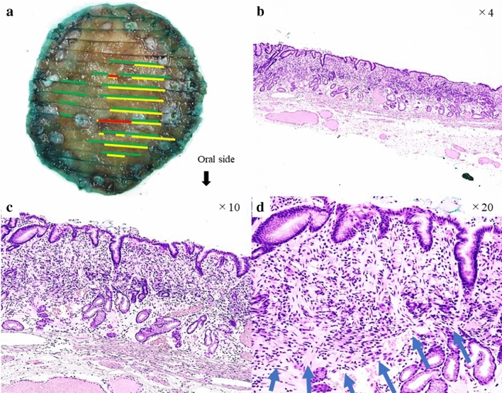
Fig. 2.
a Histopathological mapping of the resected specimen. The yellow line indicates signet ring cell carcinoma (SRCC). The red line indicates SRCC with poorly differentiated adenocarcinoma (PDA). The green line indicates fibromuscular obliteration strong positive. b Hematoxylin and Eosin staining showed that the SRCC was in the lamina propria layer and grew into the lumen forming an elevated lesion that was covered by normal mucosa. c, d The slightly depressed area at the top of the lesion contained a higher density of tumor cells compared with the surrounding area and was associated with PDA component, which was also covered by normal mucosa. The blue arrow indicates dense cancer cell growth and coexistent desmoplastic and fibrotic reactions

