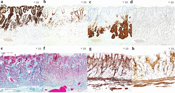
Fig. 3.
a The SRCC area and foveolar epithelium, spread over the superficial layer, were MUC5AC-positive. b The deep PDA area, extending to the middle intramucosal layer, was Ki-67-positive. c A part of the PDA area and pyloric glands and mucous neck cells, spread in the deep layer, were MUC6-positive. d A part of the PDA area was p53 positive; however, very few SRCC cells were p53-positive. e Masson trichrome staining revealed extensive fibromuscular obliteration. f This area was weakly fibromuscular as shown by obliteration in the Masson trichrome staining. g Anti-α-smooth muscle actin staining revealed fibromuscular obliteration. h This area was weakly fibromuscular as shown by obliteration in anti-α-smooth muscle actin staining

