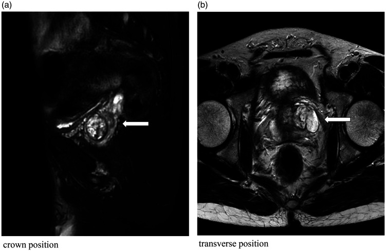
Figure 1.
Magnetic resonance images; ((a) coronal view and (b) transverse view): The prostatic volume is increased above normal, and the images show mixed signals dominated by slightly high-intensity signals surrounded by a low-signal-intensity capsule. No obviously abnormal signals are evident in the remaining prostatic tissue, and the prostatic capsule is intact (arrows).

