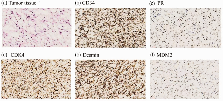
Figure 2.
Pathological and immunohistochemical findings; (a–f) Biopsy-obtained pathological sections (×400) showing heterogeneous round and spindle-shaped cells in a mucus background. Positive staining for (b) CD34, (c) PR, (d) CDK4, (e) desmin, and (f) MDM2 is widespread among the tumor cells. Staining in panel (a): hematoxylin and eosin. CD34, cluster of differentiation 34; PR, progesterone receptor; CDK4, cyclin D-dependent kinase 4.

