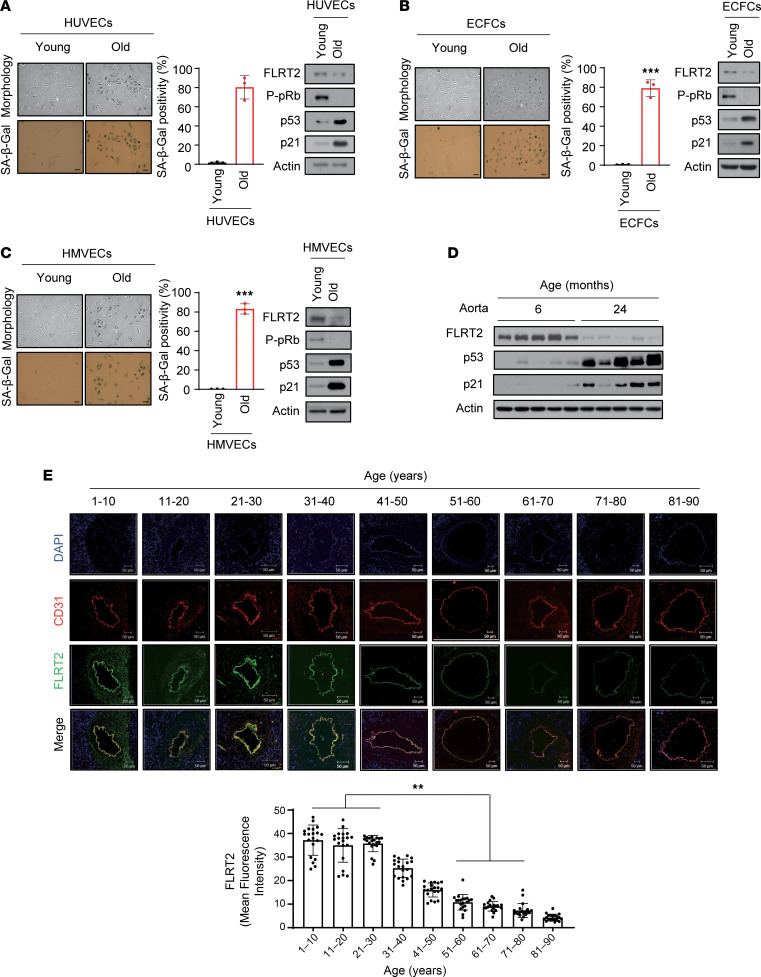
Figure 1. Lower FLRT2 expression levels in senescent endothelial cells and aged rat and human aortic tissues.
(A–C) Young (passage 4) and old (passage 15) human umbilical vein endothelial cells (HUVECs) (A), endothelial colony-forming cells (ECFCs) (B), and human microvascular endothelial cells (HMVECs) (C) were subjected to senescence-associated β-galactosidase (SA-β-Gal) and immunoblot analyses; SA-β-Gal–positive cells were quantified. Scale bar: 10 μm. The values represent mean ± SD (n = 3; ***P < 0.001). (D) Aortas of 6- and 24-month-old rats were subjected to immunoblot assays. (E) Changes in the expression levels of CD31 and FLRT2 in human arterial tissues of each age group (n = 20) were detected using immunofluorescence. Scale bar: 50 μm. The values represent mean ± SD (n = 20; **P < 0.01). Two-tailed t test.

