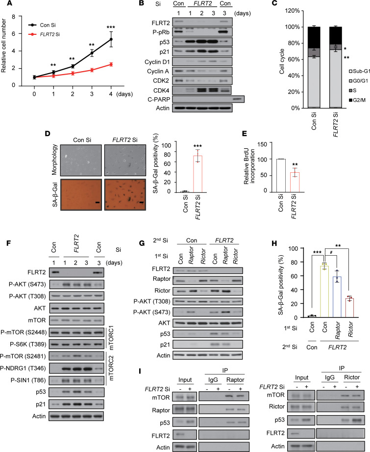
Figure 2. FLRT2 depletion induces cellular senescence through the regulation of mTORC2 in endothelial cells.
(A–F) HUVECs were transfected with 50 nM of control siRNA (Con Si) or FLRT2 siRNA (FLRT2 Si). (A) The numbers of viable cells were determined at the indicated days after transfection. (B) The cells were harvested at the indicated time points after transfection and subjected to immunoblot analysis. (C) Cell cycle distributions were analyzed using flow cytometry at day 3 after transfection. (D) Cell morphology and SA-β-Gal activity were assessed at day 3 after transfection, and the percentage of senescent cells was quantified. Scale bar: 10 μm. (E) The DNA synthesis rates of siRNA-transfected HUVECs were measured using the 5-bromo-2′-deoxy-uridine (BrdU) incorporation assay. (F) Whole-cell lysates were prepared from HUVECs transfected with Con Si and FLRT2 Si at the indicated days after transfection and subjected to immunoblot assays. (G and H) HUVECs were transfected with Con Si, Raptor Si, or Rictor Si (1st transfection) 6 hours before transfection with Con Si or FLRT2 Si (2nd transfection). At day 2 after transfection, the cells were harvested and subjected to immunoblotting using the indicated antibodies (G). SA-β-Gal activity was measured at day 3 after transfection (H). (I) HUVECs were transfected with Con Si or FLRT2 Si. At day 2 after transfection, cell lysates were subjected to immunoprecipitation with antibodies recognizing Raptor or Rictor, and immunoblot assays were performed with the indicated antibodies. The values represent mean ± SD (n = 3; #P > 0.05; *P < 0.05; **P < 0.01; ***P < 0.001). Two-tailed t test (A, D, and E), 1-way ANOVA (H).

