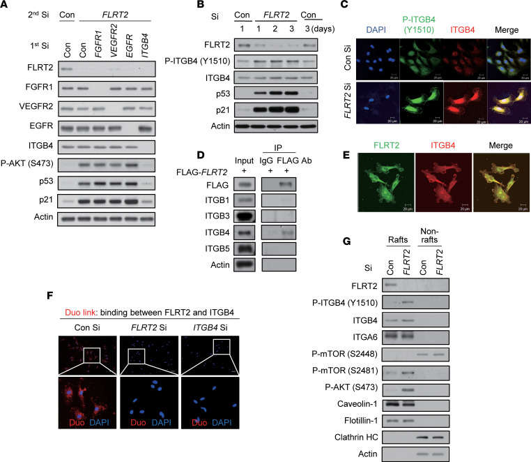
Figure 3. FLRT2 specifically binds ITGB4 and regulates ITGB4 phosphorylation in lipid rafts of the plasma membrane.
(A) HUVECs were transfected with Con Si, FGFR1 Si, VEGFR2 Si, EGFR Si, or ITGB4 Si (first transfection) 6 hours before transfection with Con Si or FLRT2 Si (second transfection). At day 2 after transfection, the cells were harvested and subjected to immunoblotting using the indicated antibodies. (B) Immunoblot assay was performed at the indicated time points after transfection of Con Si or FLRT2 Si. (C) Immunocytochemical staining was performed at day 2 after transfection of Con Si or FLRT2 Si. Scale bar: 20 μm. (D) HUVECs were transfected with a FLAG-tagged, FLRT2-overexpressing construct. At day 2 after transfection, cell lysates were subjected to immunoprecipitation with anti-FLAG antibody and immunoblotted with the indicated antibodies. (E) Immunofluorescence of nonpermeabilized HUVECs that were stained with antibodies recognizing FLRT2 or ITGB4. Scale bar: 20 μm. (F) Representative images of individual immunofluorescence staining of FLRT2 and ITGB4 interaction in HUVECs by Duo-link assay. The red dots (FLRT2/ITGB4 interaction) indicate direct interaction. Original magnification, ×40 (top) and ×200 (bottom). Scale bar: 10 μm. (G) HUVECs were transfected with Con Si or FLRT2 Si and incubated for 2 days. The lipid rafts were fractionated. Equal volumes of lipid rafts (Rafts; fractions 6–8) and nonlipid rafts (Nonrafts; fractions 2–4) were separated by SDS-PAGE and immunoblotted with the indicated antibodies.

