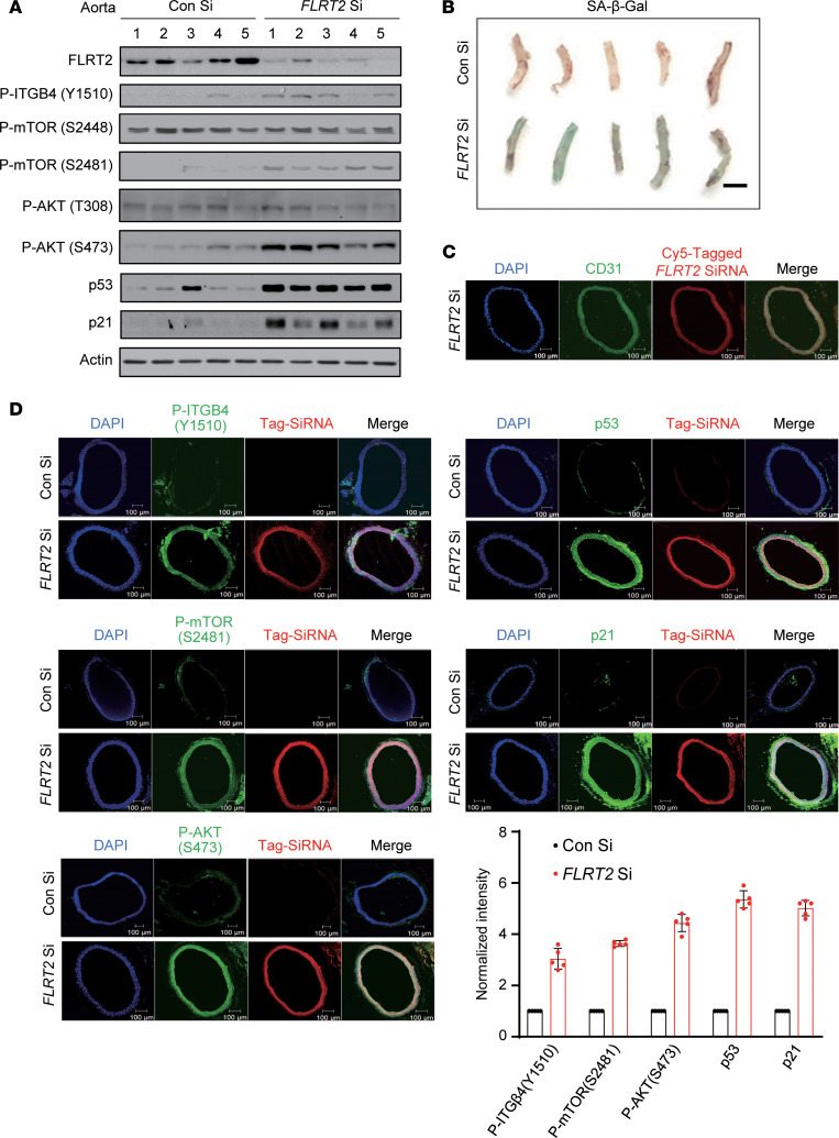
Figure 5. FLRT2 silencing promotes aortic endothelial cell senescence.
(A) Representative immunoblot images obtained from aortic lysates of mice injected intravenously with Con Si or FLRT2 Si (n = 5). (B) Images of mouse aortas assayed for SA-β-Gal activity. Scale bar: 2 mm. (C) Fluorescence micrographs of aortic cross sections obtained from mice injected intravenously with Cy5-tagged FLRT2 Si. Nuclei are stained with DAPI (blue). Red and green signals show Cy5-tagged FLRT2 Si and the endothelial marker CD31, respectively. Merged image indicates siRNA uptake by the endothelium. Scale bar: 100 μm. (D) Immunostaining for p-ITGB4 (Y1510), p-mTOR (S2481), p-AKT (S473), p53, and p21 in aortas obtained from mice injected with siRNAs. Scale bar: 100 μm. Quantitative data are shown in the graph.

