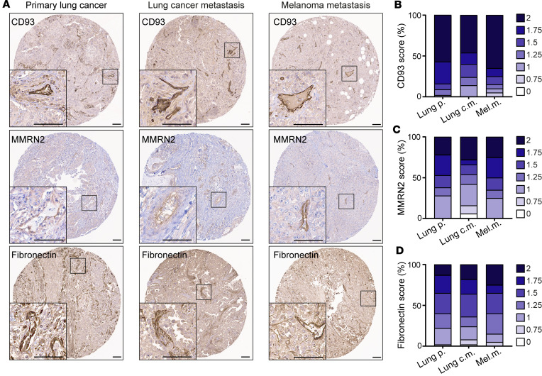
Figure 1. CD93, MMRN2, and fibronectin are highly expressed in the blood vessels of primary tumors and metastases.
(A) Immunohistochemical staining of CD93, MMRN2, and fibronectin in human tissue microarrays of primary lung cancer (n = 60), metastases originating from lung cancer (n = 50), and melanoma metastases (n = 20). Scale bars: 100 μm. Graphs represent the average of a semiquantitative scoring of CD93+ (B), MMRN2+ (C), and fibronectin+ (D) vessels performed in tumor cores of each patient by 2 researchers in a blinded fashion on a scale of 0 to 2 (0 = no vessel staining, 1 = medium intensity, and 2 = high intensity). Lung p., lung primary tumors; Lung c.m., lung cancer metastases; Mel.m., melanoma metastases.

