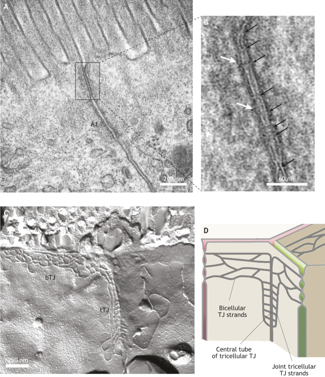
Fig. 1.
The ultrastructure of TJs. (A) Transmission electron micrograph of the AJC of mouse intestinal epithelial cells (within the jejunum), highlighting the position of the apical TJ and the neighboring AJ. (B) Detailed image of the TJ region highlighted in A, with sites of intimate plasma membrane apposition indicated by black arrows, and the electron-dense cytoplasmic plaque immediately beneath the plasma membrane indicated by white arrows. (C) FFEM image of the apical regions of epithelial intestinal (human jejunum) cells, showing the strands of a bTJ and a tTJ. (D) Cartoon depicting bTJ and tTJ structure, highlighting the positions of bTJ strands, joint tTJ strands and the central tube of a tTJ. Images in A and B provided by Kyoko Furuse, National Institute for Physiological Sciences, Okazaki, Japan. Image in C provided by Susanne M. Krug, Charité – Universitätsmedizin Berlin, Germany.

