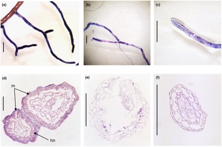
FIGURE B1.

Stained Leptospermum root tips under a dissecting microscope (a–c) and corresponding cross sections under a compound microscope at 40 × magnification (d–f). (d) Shows a mantle sheath (m) and Hartig Net (hn) present in the roots of (a). No ectomycorrhizal structures were visible in cross sections of the roots of (b and c) (e and f). Scales bars are approximately 1 mm in A to C and 50 μm in D to F.
