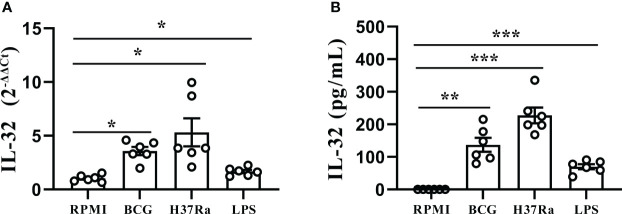
Figure 4.
Stimulation of IL-32 by Mtb stimulus. (A) PFMCs isolated from fresh TPE were stimulated with RPMI, LPS, BCG, and H37Ra for 24 hours. IL-32 mRNA was measured by quantitative PCR and normalized against the housekeeping gene GAPDH, and the expression was calculated as 2(−ΔΔCt) (mean fold change ± SEM, n = 6). (B) PFMCs were stimulated with RPMI, LPS, BCG, or H37Ra; Triton X-100 (0.5%) was added 24 hours later; IL-32 concentration was measured by ELISA. Data are presented as means ± SEM (n = 6). *p < 0.05; **p < 0.01; ***p < 0.001. PFMCs, pleural fluid mononuclear cells; TPE, tuberculous pleural effusion; RPMI, Roswell Park Memorial Institute; LPS, lipopolysaccharide; BCG, Bacillus Calmette-Guérin.

