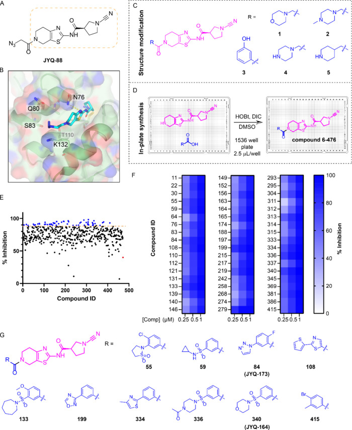
Figure 1.
Discovery of improved PARK7 inhibitors. (A) Structure of PARK7 inhibitor JYQ-88. (B) Crystal structure of PARK7-JYQ-88 complex (PDB 7PA3) showing an unoccupied pocket around the azidoacetyl moiety. (C) Chemical structures of designed compounds with a hydrophilic substituent. (D) Schematic illustration of in-plate synthesis to build a compound library. (E) Screening results using an FP assay at 1 μM compound concentration. Blue color represents compounds showing over 90% inhibition. Red color shows inhibition data for the amine precursor compound. (F) Heatmap displaying validation of the screening hits at 0.25, 0.5, and 1 μM using the FP assay. (G) Chemical structures of the resynthesized top 10 compounds.

