Figure 3.
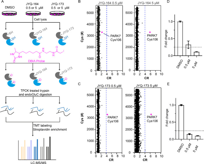
Investigation of the cellular selectivities of JYQ-164 and JYQ-173. (A) Schematic illustration of the workflow for the SLC-ABPP experiment. (B, C) SLC-ABPP profiling of PARK7 inhibitor JYQ-164 or JYQ-173 in A549 cells using two different concentrations (0.5 and 5 μM) quantified >5500 cysteine sites. All experiments were performed in triplicates. Data are represented as means ± s.d. Dotted lines represent a CR threshold of 4 (75% reduction in DBIA probe binding). (D, E) Fold changes of annotated peptide for PARK7 Cys106 residue in inhibitor-treated samples compared to a DMSO control are represented as a column graphic where the 0.25 fold change corresponding to 75% reduction in DBIA probe binding is highlighted with dotted lines.

