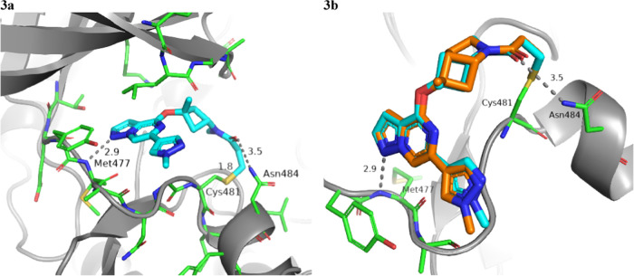
Figure 3.
(a) X-ray cocrystal structure and binding mode of 25 (cyan; PDB 8TU4) in the active site of BTK. Hydrogen bond interactions are shown as gray dotted lines. (b) Superposition of the binding modes of compound 25 (cyan) and compound 27 (orange; PDB 8TU5) in the BTK ATP binding site was done from a top-down view with Met477 to the bottom left and Asn848 to the right (Cys481 in the back).

