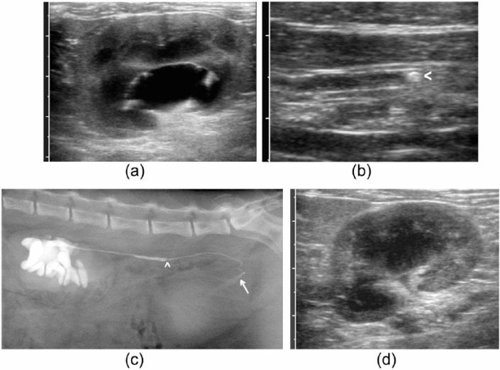
Figure 2.
Example of a cat with pelvic dilatation and ureteral calculus compatible with ureteral obstruction but patent ureter on pyelography. (a) Dorsal plane ultrasound image of the left kidney at presentation. Pelvic diameter was recorded as 10 mm; (b) dilated left ureter containing a calculus (arrowhead); (c) lateral radiograph after pyelography showing localised dilatation of the left ureter cranial to an intraluminal filling defect compatible with a calculus (arrowhead) and passage of contrast into the bladder (arrow); (d) dorsal plane ultrasound image of the left kidney at follow-up examination after medical management for 1 month, in which pelvic dilatation has resolved

