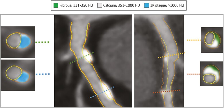
Figure 3.
Example of 1K plaque. The artery segment in the left panel (A) shows two lesions composed of 1K plaque without non-calcified plaque. Cross-sectional examples are shown with 1K plaque. The artery segment in (B) shows calcifications between 351 and 1000 HU intermingled in non-calcified plaque. Two cross-sections show 351 to 1000 HU calcium together with fibrous plaque tissue. HU, Hounsfield units. Adapted from van Rosendael et al.92

