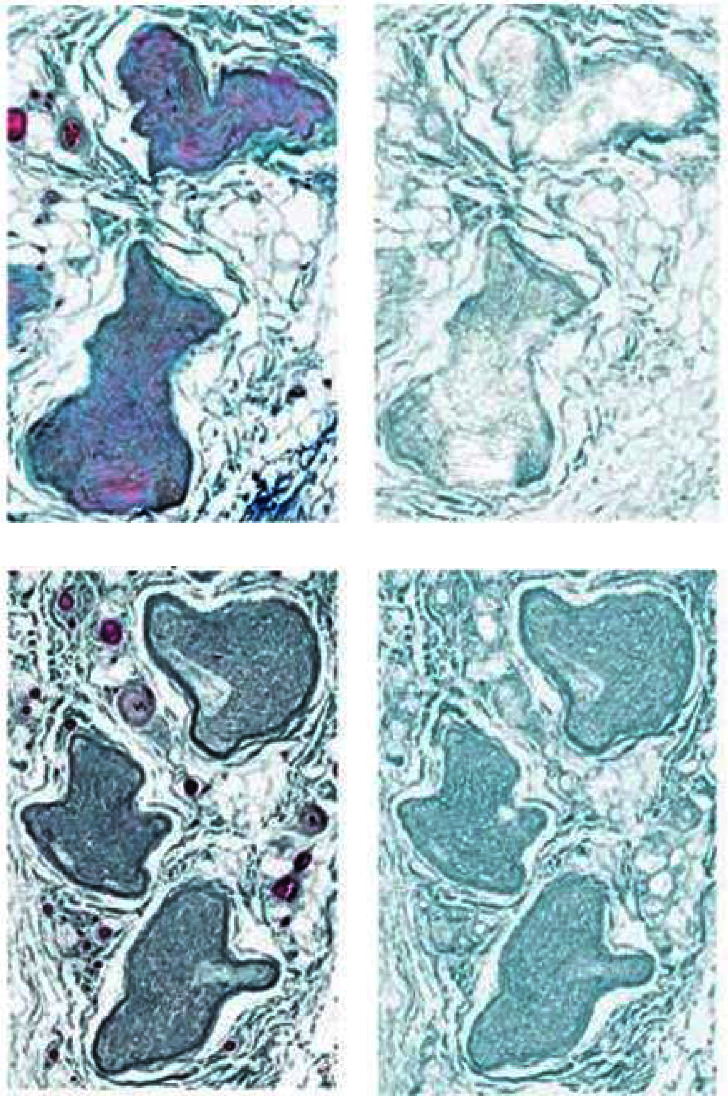
Figure 2.

Representative protected and affected ilioinguinal nerve samples stained with trichrome (a, c), next to the resultant color deconvoluted image containing only the green component (b, d), showing increased collagen content in the inguinal canal sample. (a) Proximal sample. (b) Proximal green component. (c) Inguinal sample. (d) Inguinal green component.
