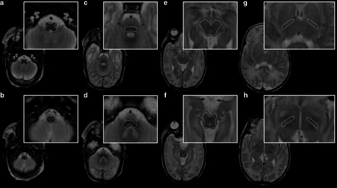
Fig. 1.
ROI drawings in a female infant (GA at birth: 27 + 5; MRI at 35 + 6) on a SyMRI-based, T2-weighted MR contrast (repetition time (TR)/echo time (TE): 4500/100 ms). Wherever applicable, two separate measurements were performed at different levels for each brain area of interest: e.g., medulla oblongata (a level of inferior olivary nucleus; b level of gracile and cuneate nucleus); pontine tegmentum (c level of superior olive; d level of the pontine vestibular nuclei); midbrain (e level of superior colliculi/intercollicular section; f level of inferior colliculi); and PLIC (g level of the interventricular foramen; h level of the third ventricle)

